Skip to main content
Wellcome Collection homepage
Visit us
What’s on
Stories
Collections
Get involved
About us
Sign in to your library account
Search for anything
Library account
Search for anything
Search
Home
|
Collections
Prosencephalon
Forward-most part of the brain
Wikidata
Source:
Wikidata
On this page
On this page
Images from the collections
Works from the collections
Related topics
Images from the collections
Images referencing Prosencephalon
13 images from works
View all
Works from the collections
13 works
Digital Images
Online
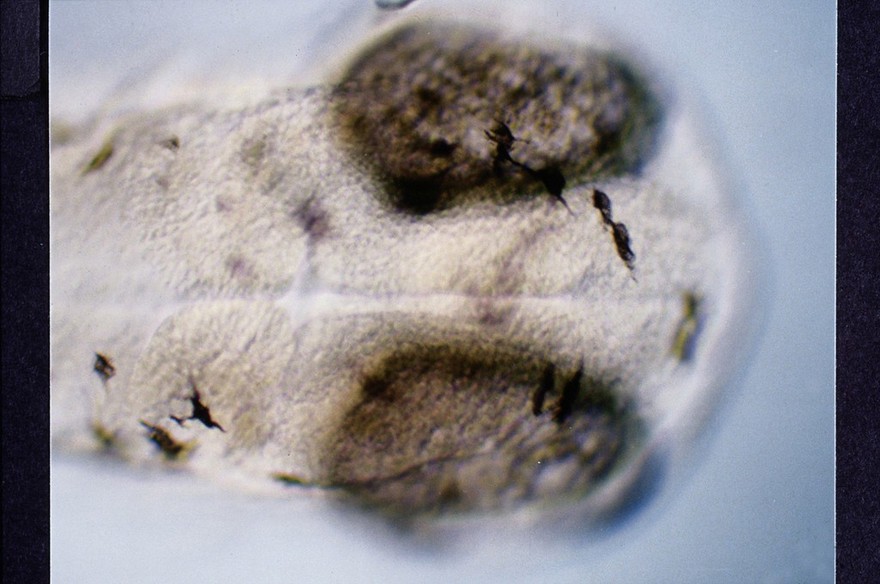
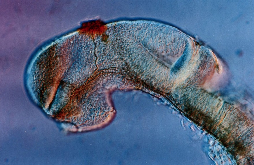
Zebrafish eye development
Dr Steve Wilson
Digital Images
Online
Zebrafish eye development
Dr Steve Wilson
Digital Images
Online
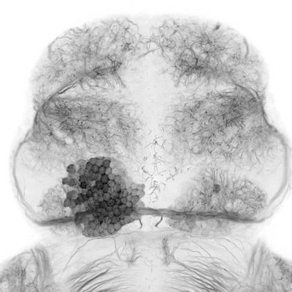
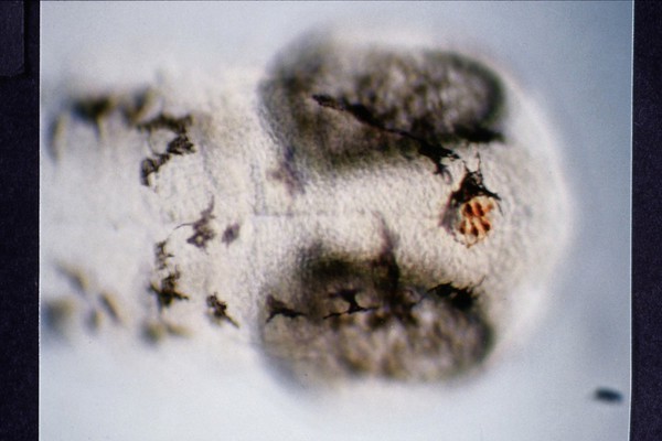
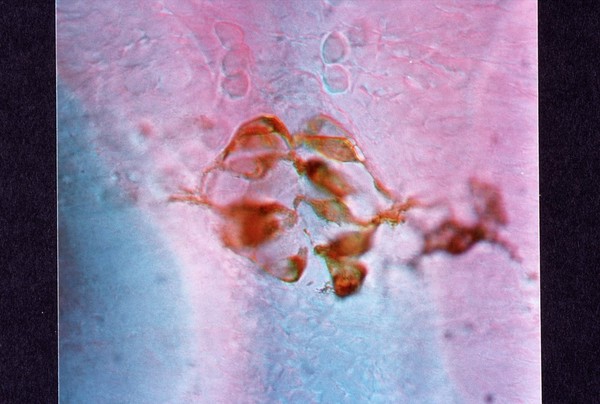
Dorsal view of the forebrain of a wild-type zebrafish embryo
Ana Faro, Dr Steve Wilson
Digital Images
Online
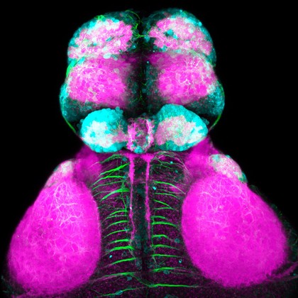
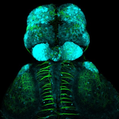
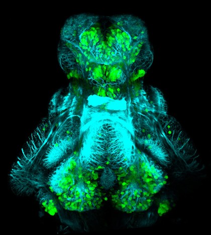
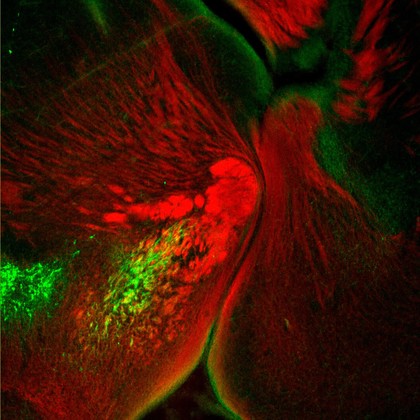
Dopaminergic neurons in the zebrafish forebrain. Confocal micrograph of a 4 day old transgenic zebrafish embryo viewed from a lateral aspect. Neurons in the olfactory bulb, telencepahlon, ventral diencephalon, pretectum and hypothalamus are labelled in green. Axonal tracts are shown in cyan and neuropil in magenta. In order to show the anatomy of the brain better the skin and eyes of the embryo have been removed post-fixation.
Kate Turner, Dr Steve Wilson
Digital Images
Online
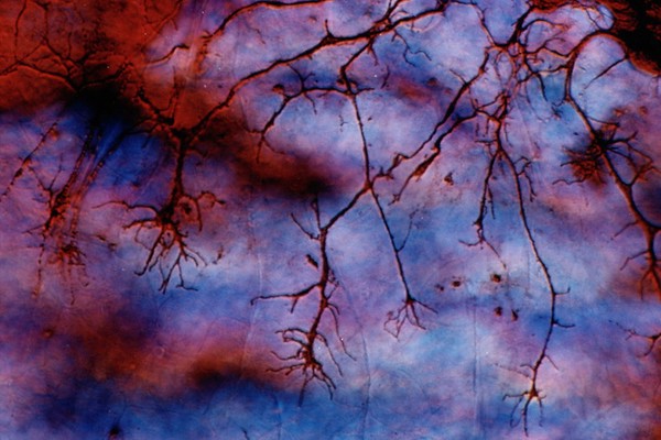
Zebrafish eye development
Dr Steve Wilson
View all
Related topics
Development
Embryonic Structures
PHOTORECEPTOR
Model organism
Aquatic vertebrae
CNS
Epiphyses
Neural network
Neuronal signalling
Cyan
Close modal window