Hermann Aberle, University of Munster
Images from the collections
Images by Hermann Aberle, University of Munster
9 images from works


Works from the collections
9 works

- Digital Images
- Online
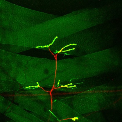
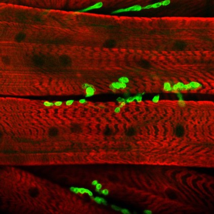
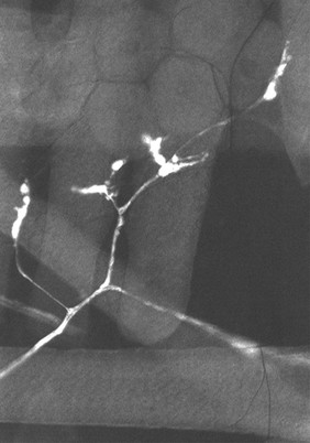
Muscle Innervation
Hermann Aberle, University of Munster
- Digital Images
- Online
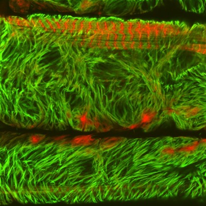
Muscle Innervation in Drosophila
Hermann Aberle, University of Munster
- Digital Images
- Online
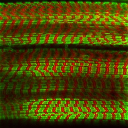
Mutant Muscle Sarcomere, Drosophila larva
Hermann Aberle, University of Munster
- Digital Images
- Online
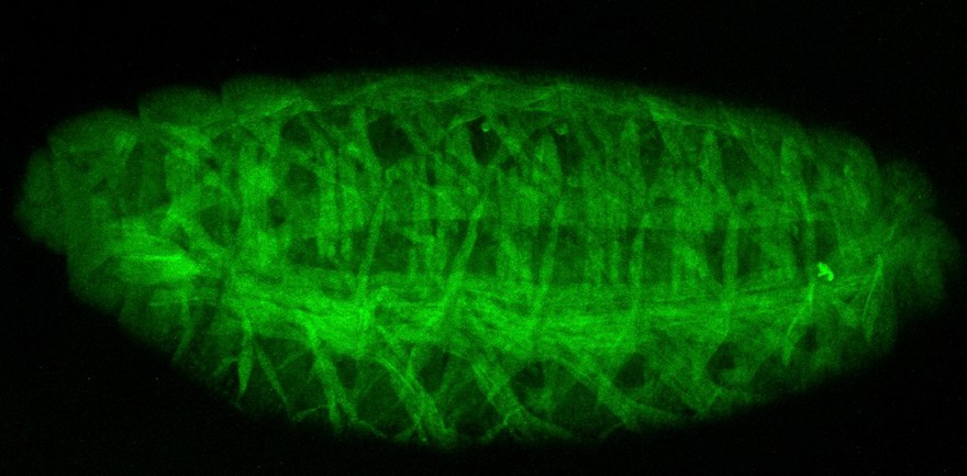
Muscle fibres, Drosophila embryo
Hermann Aberle, University of Munster
- Digital Images
- Online
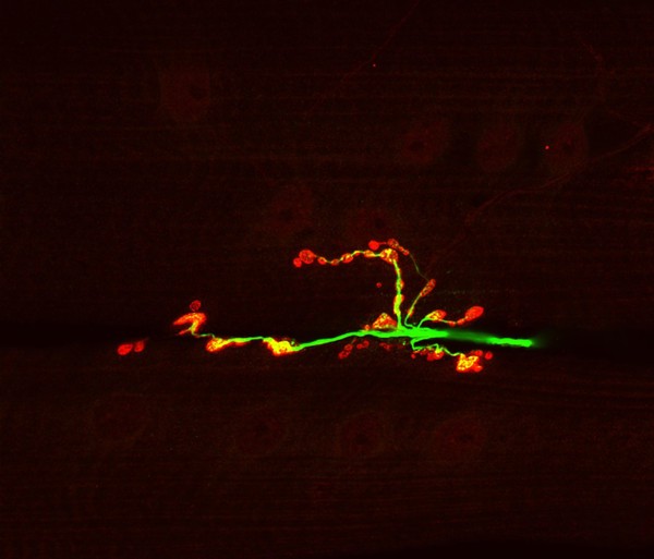
Neuromuscular junction from a Drosophila larva showing a nerve synapse atttached to a muscle fibre. The transmission of signals in shown via synaptic vesicles containing neurotransmitter substance (shown in red). These vesicles are in close accosiation with the microtubule-based synaptic core cytoskeleton o the nerve, which is shown in green.
Hermann Aberle, University of Munster