Villus
Images from the collections
Images referencing Villus
11 images from works

Works from the collections
11 works

- Digital Images
- Online
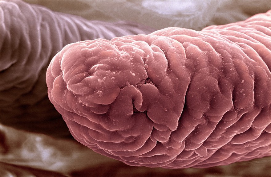
Intestinal villi
Liz Hirst, Medical Research Council
- Digital Images
- Online
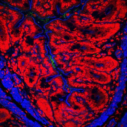
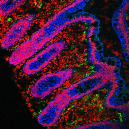
Human small intestine showing villi and glands. The cytokeratinin the cells is stained blue, the cell nuclei are stained red and the endothelial cells lining the blood vessels are stained green.
S. Schuller
- Digital Images
- Online
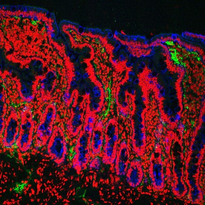
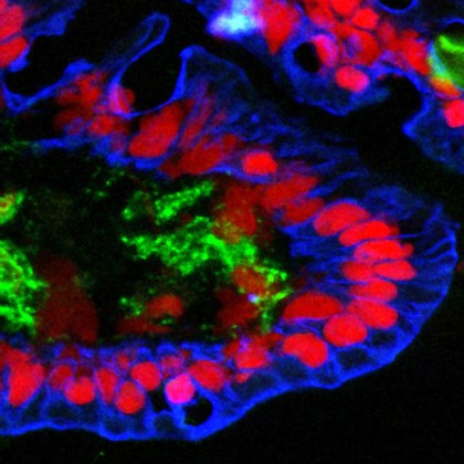
Mouse colon infected with Citrobacter rodentium
S. Schuller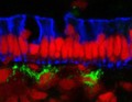
- Digital Images
- Online
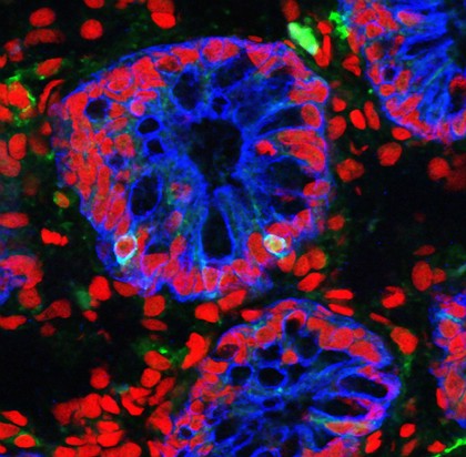
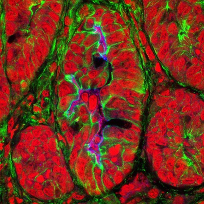
Human small intestine showing the columnar epithelium. The cytokeratinin the cells is stained blue, the cell nuclei are stained red and the endothelial cells lining the blood vessels are stained green.
S. Schuller
- Digital Images
- Online
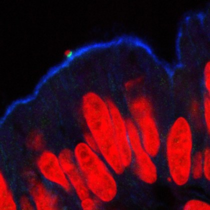
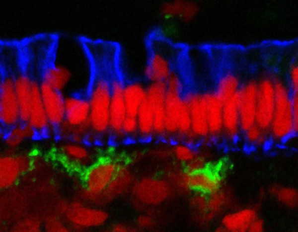
A single enteropathogenic E. coli in the intestine
S. Schuller