Wellcome Collection homepage
  • Visit us
  • What’s on
  • Stories
  • Collections
  • Get involved
  • About us
Sign in to your library account
Search for anything
Library account
Home| Collections

Human anatomy

Part of
Anatomy
Medical sciences
Human biology

On this page

  • Images from the collections
  • Works from the collections
  • Related topics

Images from the collections

Images about Human anatomy

1,855 images from works
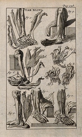
  • Muscles of the lower leg and foot. Engraving, 1686, after Gérard de Lairesse, 1685.
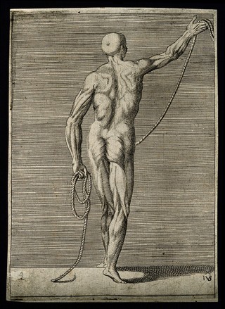
  • An écorché figure seen from the back, holding a length of rope. Engraving by G. Bonasone, 155-.
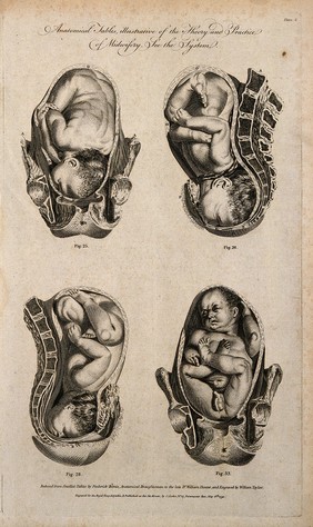
  • Foetuses in utero: four figures showing cross-sections of a foetus in various positions in the uterus. Line engraving, 1791, by W. Taylor after F. Birnie, after W. Smellie.
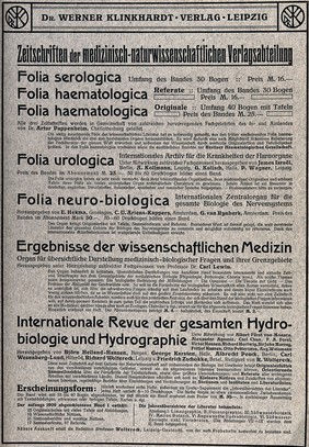
  • List of medical periodicals, including prices, printed on verso of contents page to Alfred Kast, Pathologisch-anatomische Tafeln. Letterpress, ca. 1897.
  • M0018670EB: Anatomical male figure in ivory
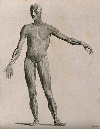
  • An écorché with left arm extended to the side, seen from the front. Line engraving by Campbell, 1816/1821.
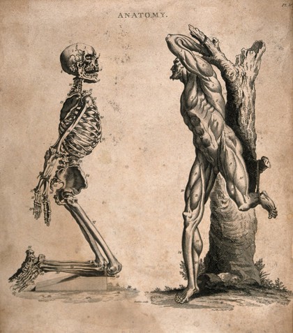
  • Two figures: a kneeling human skeleton, with hands tied behind its back, and an écorché figure tied to a tree-trunk. Engraving by Barlow, ca. 1800.
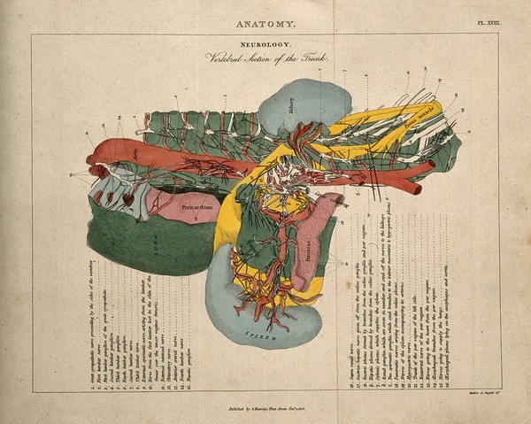
  • Torso: cross-section indicating the nerves, organs, arteries and bones, in various colours. Coloured line engraving by H. Mutlow, 1808.
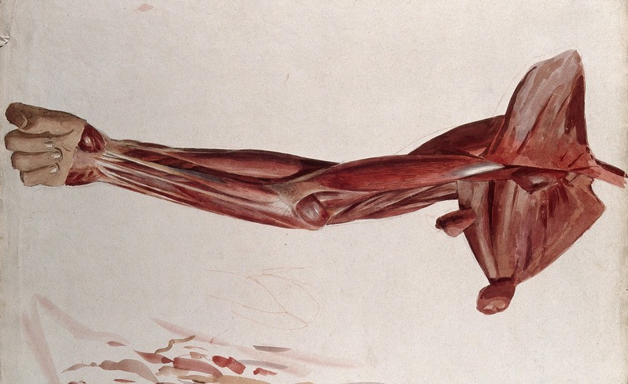
  • Dissection of the trunk: side view, showing the bones and muscles, with small pencil sketches of a nude in various poses. Ink and watercolour with laminated flaps, 18--.
  • Anatomical figures with detachable parts. Colour lithographs by American Manikin Co., ca. 1890.
View all

Works from the collections

2,454 works

    • Pictures
    • Online

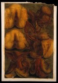
    Dissections of the female urogenital system: six figures. Colour mezzotint by J. F. Gautier d'Agoty after himself, 1754.

    | Jacques Fabien Gautier d'Agoty | Date: [1754] | Reference: 572095i
    • Pictures

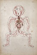
    The arteries of the human body with a foetus in the womb. Watercolour by a Persian artist.

    | Date: [between 1800 and 1899?] | Reference: 582965i
    • Books
    • Online

    The anatomy of the thymus gland / by Sir Astley Cooper.

    | Astley Cooper | Date: 1832
    • Pictures

    Diagrams of the human torso, left and right side views, with internal organs indicated. Colour lithograph by F. Reichhold, 1890/1910?.

    | Reichhold, F. | Date: [1890/1910?] | Reference: 569387i
    • Pictures
    • Online

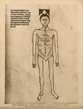
    A male nude with the parts of the abdomen and thorax labelled. Colour process print, 1926, after a manuscript illustration, 1345.

    | Date: [1926] | Reference: 26656i
View all

Related topics

Muscles
Anatomy
Brain
Blood Circulation
Nervous System
Models, Anatomic
Human dissection
Surgical Instruments
Human skeleton
Bone and Bones

Wellcome Collection

183 Euston Road
London NW1 2BE

+44 (0)20 7611 2222
info@wellcomecollection.org

  • Getting here

Today’s opening times

  • Galleries
    10:00 – 18:00
  • Library
    10:00 – 18:00
  • Café
    10:00 – 18:00
  • Shop
    10:00 – 18:00

Opening times

Our building has:

  • Step free access
  • Hearing loops

Accessibility

  • Visit us
  • What’s on
  • Stories
  • Collections
  • Get involved
  • About us
  • Contact us
  • Jobs
  • Media office
  • Developers
  • Privacy and terms
  • Cookie policy
  • Manage cookies
  • Modern slavery statement
TikTok
Facebook
Instagram
YouTube

Except where otherwise noted, content on this site is licensed under a Creative Commons Attribution 4.0 International Licence