Notocord
Images from the collections
Images referencing Notocord
5 images from works
Works from the collections
5 works
- Digital Images
- Online
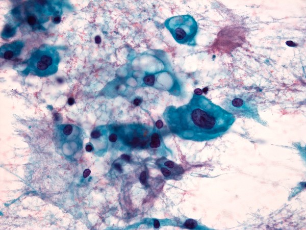
Papanicolaou stained smear of a clival chordoma, microscopy. Chordomas are cancers formed of cells which resemble those of the notochord (spine) of a developing foetus. Although they can present anywhere within the spine and skull, the majority grow in the sacral region of the spine, corresponding to the lower back. This image shows a Papanicolaou (Pap) stained smear obtained from a needle biopsy of a chordoma in the clivus, a part of the cranium at the base of the skull.
William R. Geddie
- Digital Images
- Online
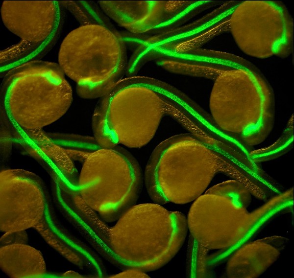
Zebrafish embryos with green fluorescent notocords
S. Roy & F. Muller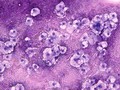
- Digital Images
- Online
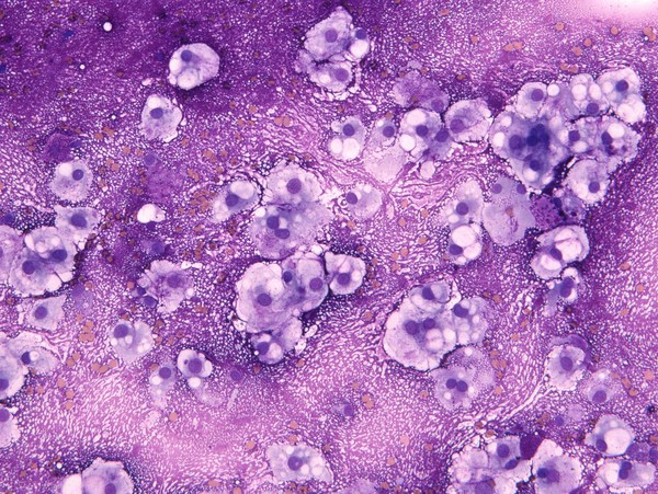
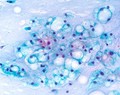
MGG stained smear of a C2 vertebral chordomal mass
William R. Geddie
- Digital Images
- Online
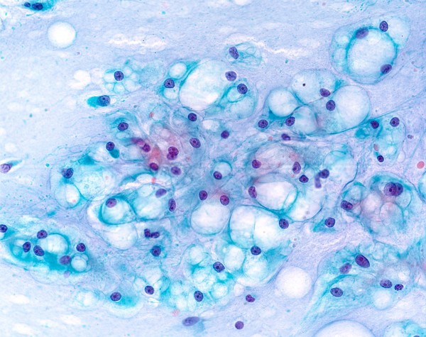
Papanicolaou stained smear of a C2 vertebral chordomal mass, microscopy. Chordomas are cancers formed of cells which resemble those of the notochord (spine) of a developing foetus. Although they can present anywhere within the spine and skull, the majority grow in the sacral region of the spine, corresponding to the lower back. This image shows a Papanicolaou (pap) stained smear obtained from a needle biopsy of a chordoma of the C2 vertebrae, located at the top of the neck just underneath the base of the skull.
William R. Geddie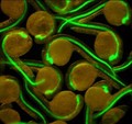
- Digital Images
- Online
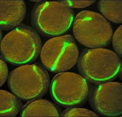
Zebrafish embryos with green fluorescent midlines
S. Roy & F. Muller