Immunology
Images from the collections


Works from the collections
82 works
- Digital Images
- Online
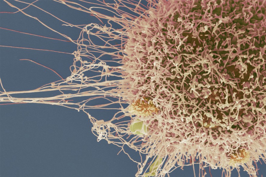
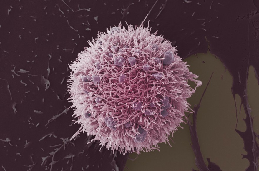
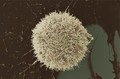
HeLa cell, immortal human epithelial cancer cell line, SEM
Anne Weston, Francis Crick Institute
- Digital Images
- Online
Trichuris muris is a parasitic nematode affecting mice. Following ingestion, T. muris eggs hatch in the large intestine where they develop into adults. The anterior end of the worm burrows into the lining of the gut, leaving the posterior end protruding into the lumen of the gut. The worms mate in this orientation, and the resulting eggs are released in to the gut lumen and shed faecally.
David Goulding, Wellcome Trust Sanger Institute
- Digital Images
- Online
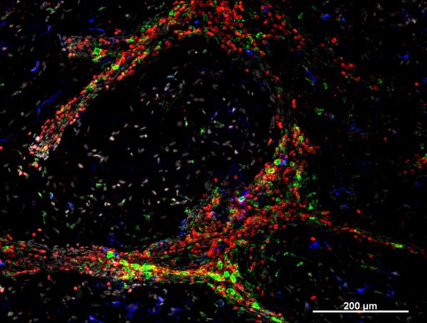
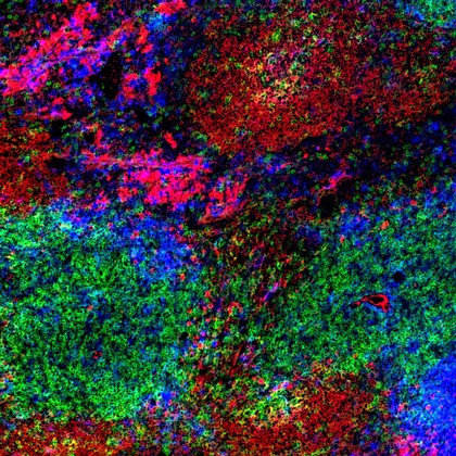
Normal spleen showing B cells and T cells
Peter Lane and Fiona McConnell
- Digital Images
- Online
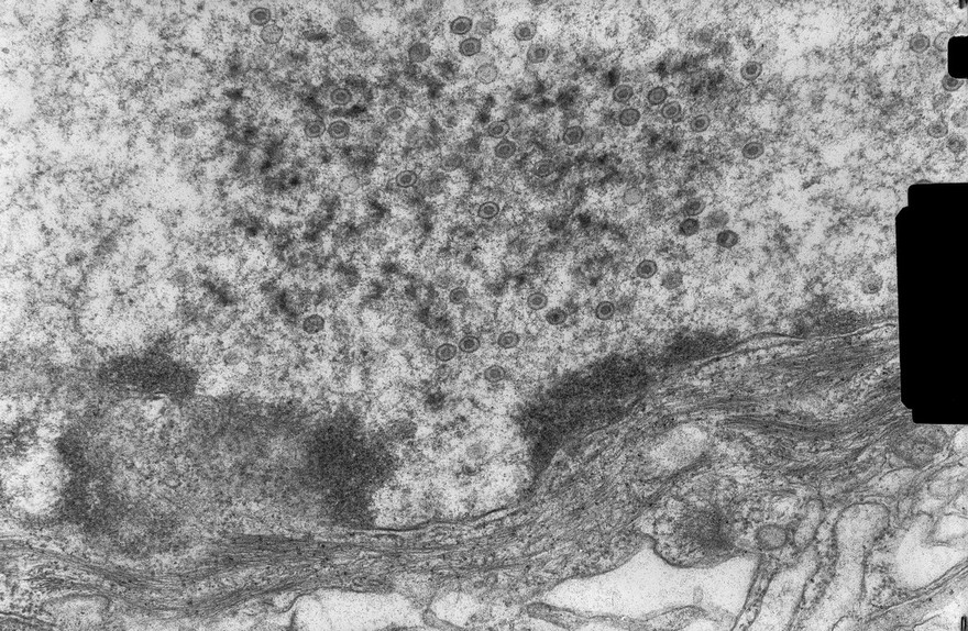
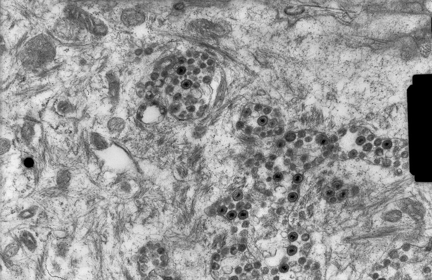
Lymphocyte with mitochondria
Rob Young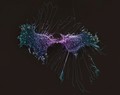
- Digital Images
- Online
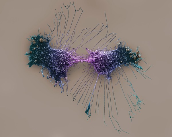
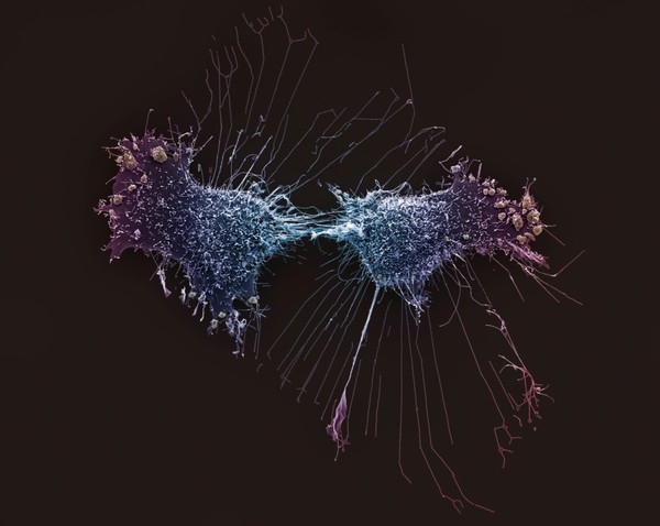
HeLa cells, immortal human epithelial cancer cell line, SEM
Anne Weston, Francis Crick Institute