Foedisch, Friedrich, active 1875-1877.
Images from the collections
Images by Foedisch, Friedrich, active 1875-1877.
8 images from works


Works from the collections
8 works

- Pictures
- Online
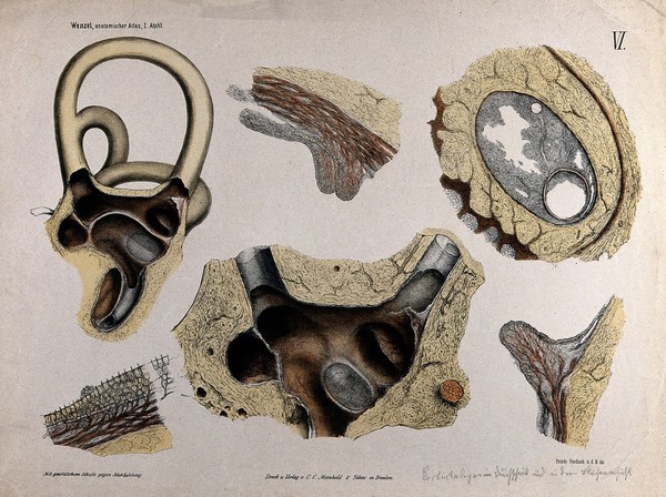
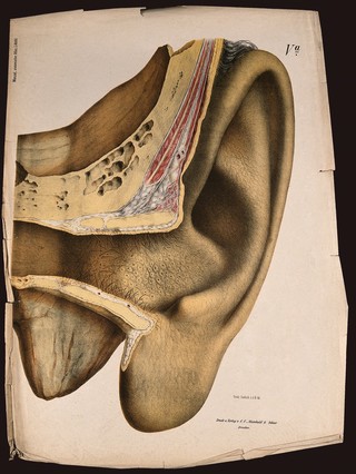
The ear: section showing the internal structure. Colour lithograph by F. Foedisch, ca. 1875.
Foedisch, Friedrich, active 1875-1877.Date: [1875?]Reference: 572781i
- Pictures
- Online
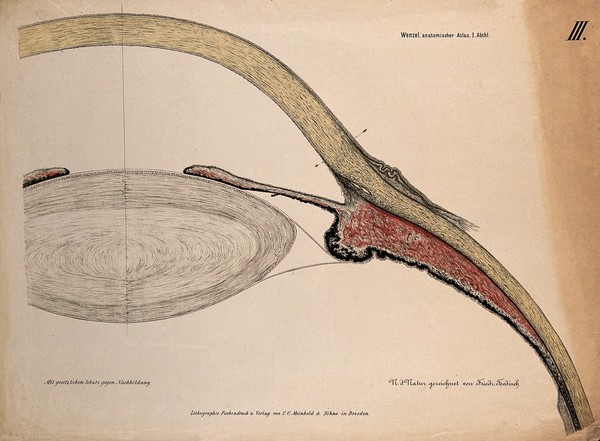
The eye: section of the ciliary body, cornea and lens. Colour lithograph by F. Foedisch, 1875.
Foedisch, Friedrich, active 1875-1877.Date: [1875]Reference: 572777i
- Pictures
- Online
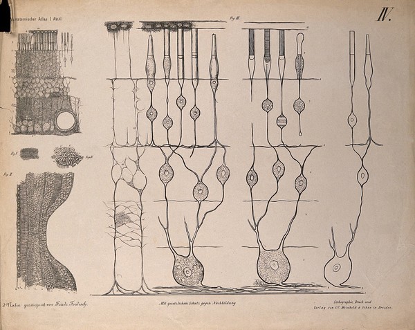
The eye: five diagrams showing the microscopic structure of the retina. Lithograph by F. Foedisch, 1875.
Foedisch, Friedrich, active 1875-1877.Date: [1875]Reference: 572778i
- Pictures
- Online
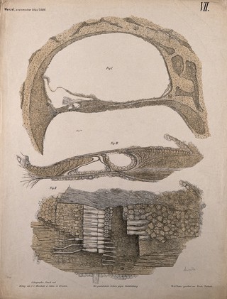
The ear: three diagrams showing the microscopic structure of ampulla of the semicircular canals. Colour lithograph by F. Foedisch, ca. 1875.
Foedisch, Friedrich, active 1875-1877.Date: [1875?]Reference: 572782i
- Pictures
- Online
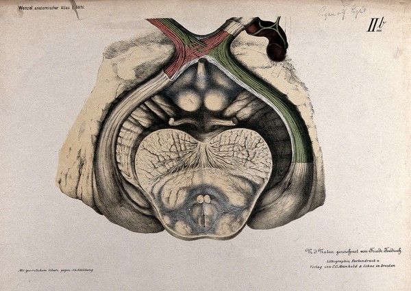
Anatomical section of the brain showing the optic chiasma. Colour lithograph by F. Foedisch, 1875.
Foedisch, Friedrich, active 1875-1877.Date: [1875]Reference: 576666i