Some uses of a conductivity cannula in the cat / by J.A.B. Grayu and W.D.M. Paton.
- Gray, J. A. B.
- Date:
- [1948?]
Licence: In copyright
Credit: Some uses of a conductivity cannula in the cat / by J.A.B. Grayu and W.D.M. Paton. Source: Wellcome Collection.
1/2
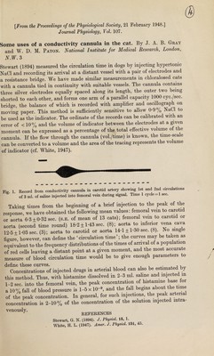
![[From the Proceedings of the Physiological Society, 21 February 1948.] Journal Physiology, FoZ. 107. Some uses of a conductivity cannula in the cat. By J. A. B. Gray and W. D. M. Paton. National Institute for Medical Research, London, N.W. 3 Stewart (1894) measured the circulation time in dogs by injecting hypertonic NaCl and recording its arrival at a distant vessel with a pair of electrodes and a resistance bridge. We have made similar measurements in chloralosed cats with a cannula tied in continuity with suitable vessels. The cannula contains three silver electrodes equally spaced along its length, the outer two being shorted to each other, and forms one arm of a parallel capacity 1000 cyc./sec. bridge, the balance of which is recorded with amplifier and oscillograph on moving paper. This method is sufficiently sensitive to allow 0-9% NaCl to be used as the indicator. The ordinate of the records can be calibrated with an error of < 10% and the volume of indicator between the electrodes at a given moment can be expressed as a percentage of the total effective volume of the cannula. If the flow through the cannula (vol./time) is known, the time-scale can be converted to a volume and the area of the tracing represents the volume of indicator (cf. White, 1947). Fig 1 Record from conductivity cannula in carotid artery showing 1st and 2nd circulations * of 3 ml. of saline injected into femoral vein during signal. Time 1 cycle = 1 sec. Taking times from the beginning of a brief injection to the peak of the response, we have obtained the following mean values: femoral vein to carotid or aorta 6-3±0-32 sec. (s.e. of mean of 13 cats); femoral vein to carotid or aorta (second time round) 18-2 ±1-43 sec. (8); aorta to inferior vena cava 12-5 ±1*03 sec. (3); aorta to carotid or aorta 14*1 ± 1*30 sec. (8). No single figure, however, can define the ‘circulation time’; the curves may be taken as equivalent to the frequency distributions of the times of arrival of a population of red cells leaving a distant point at a given moment, and the most accurate measure of blood circulation time would be to give enough parameters to define these curves. . , A Concentrations of injected drugs in arterial blood can also be estimated by this method. Thus, with histamine dissolved in 2-3 ml. saline and injected m 1-2 sec. into the femoral vein, the peak concentration of histamine base for a 10% fall of blood pressure is 1-5 x 10-». and the fall begins about the time of the peak concentration. In general, for such injections, the peak ar ena concentration is 2-10% of the concentration of the solution injected mtra- Ven0U8ly- ™,RENCES Stewart, G. N. (1894). J. Physiol. 15, 1. White, H. L. (1947). Amer. J. Physiol. 151, 45.](https://iiif.wellcomecollection.org/image/b30632614_0001.jp2/full/800%2C/0/default.jpg)