Clay and wax modelling of the living urinary bladder under electric light / by E. Hurry Fenwick.
- Fenwick, Edwin Hurry, 1856-1944.
- Date:
- 1889
Licence: Public Domain Mark
Credit: Clay and wax modelling of the living urinary bladder under electric light / by E. Hurry Fenwick. Source: Wellcome Collection.
Provider: This material has been provided by The Royal College of Surgeons of England. The original may be consulted at The Royal College of Surgeons of England.
1/4
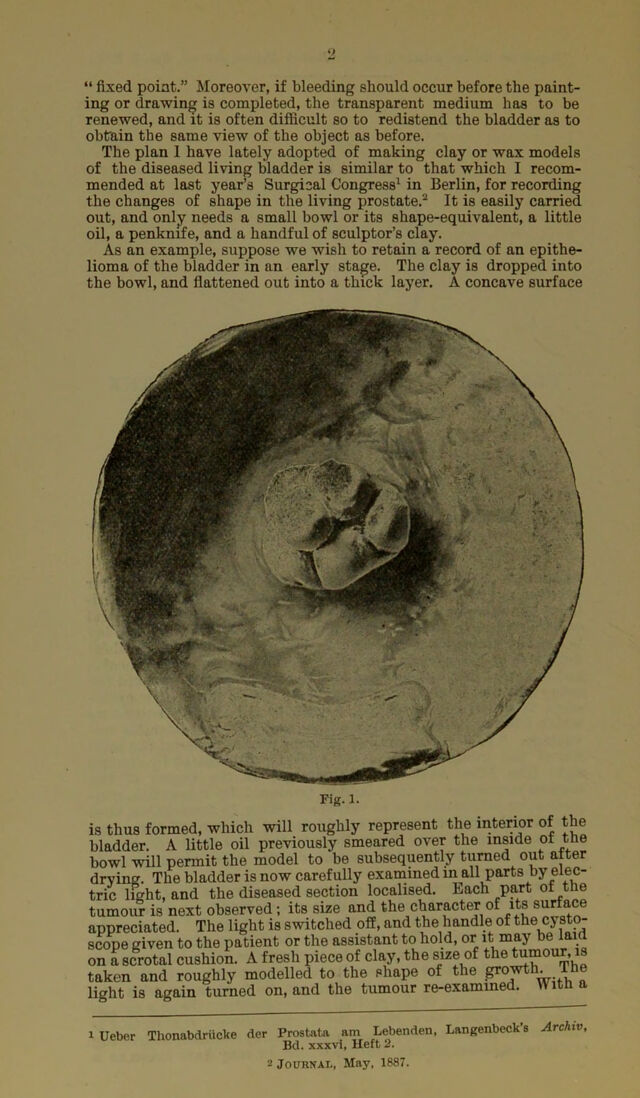
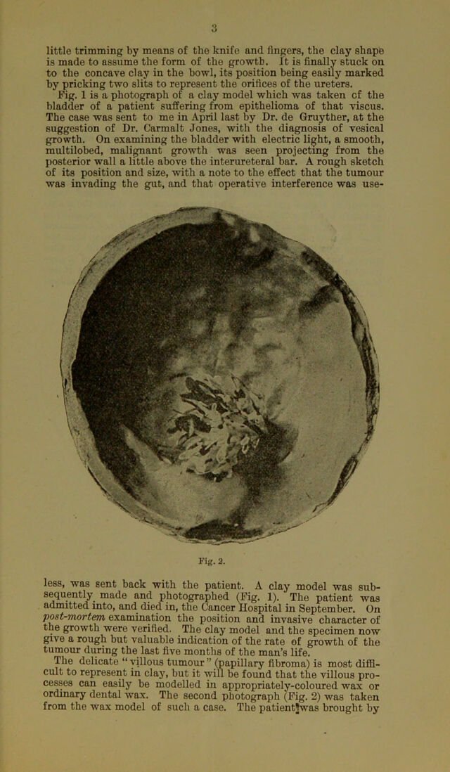
![[Keprinted for the Author from the British Medical Journal, Jan. 5,1889.] CLAY AND WAX MODELLING LIVING URINAKY BLADDER ELECTRIC LIGHT. OF THE UNDER By E. hurry FENWICK, F.R.C.S., Surgeon (Out-patient) to St. Peter’s Hospital for Urinary Disease.; Assistant- Surgeon to the London Hospital. Although Mr. Pearson-Cooper and I have been at last able, after overcoming great difficulties, to obtain good photographs of arti- ficial growths in male bladders post mortem, yet our vesical camera will have to be considerably improved before we can lay before the profession a practical apparatus for obtaining faithful negatives of the mucous membrane of the living viscus in health and disease. But, until we are able to thus graphically record the many new and interesting clinical facts which the electric cysto- scope is constantly revealing, I wish to advocate a substitute which I have employed for some time, and with considerable ad- vantage. I refer to modelling in some plastic material the inte- rior of the living bladder as it appears illuminated by electric light (the Nitze method). The changes in the aspect of the mucous membrane of the bladder, produced by relaxation, con- gestion. or infiltration are so varied and often so remarkable, that it is only by systematically accumulating a record of these ap- pearances that a sure basis for establishing a sound diagnosis, prognosis, and treatment of vesical disease upon visual grounds can be acquired. Drawings in pencil, pen, or colour are most valuable if carefully taken, but they fall far short of clay or wax modelling in convey- ing to others an exact idea of the disease depicted. The reasons for this are obvious. The cystoscopic field from which the artist draws is small, and the area to be portrayed is often large; hence a number of drawings is generally necessary to represent the dis- ease in its entirety. The cystoscopic field changes with the slightest movement of the patient or the operator. Even a cough, a deep inspiration, or a slight involuntary vesical contraction is sufficient to puzzle a non-professional artist, by suddenly displacing or distorting some salient feature which he may have taken as a](https://iiif.wellcomecollection.org/image/b22455036_0003.jp2/full/800%2C/0/default.jpg)