The effect of light on the response of frogs to drugs / by E. Boock and J.W. Trevan.
- Boock, Ellen.
- Date:
- [1923?]
Licence: In copyright
Credit: The effect of light on the response of frogs to drugs / by E. Boock and J.W. Trevan. Source: Wellcome Collection.
1/2
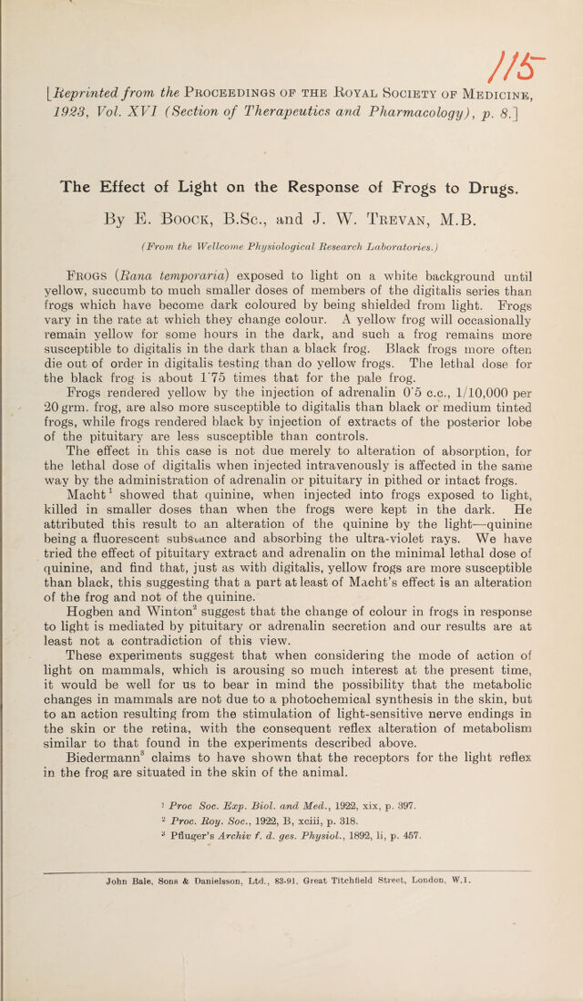
![|_Reprinted from the Proceedings of the Hoyal Society of Medicine, 1923, Vol. XVI (Section of Therapeutics and Pharmacology), p. 8.] The Effect of Light on the Response of Frogs to Drugs. By E. Boock, B.Sc., and J. W. Trevan, M.B. (From the Wellcome Physiological Research Laboratories.) FROGS (Rana temporaria) exposed to light on a white background until yellow, succumb to much smaller doses of members of the digitalis series than frogs which have become dark coloured by being shielded from light. Frogs vary in the rate at which they change colour. A yellow frog will occasionally remain yellow for some hours in the dark, and such a frog remains more susceptible to digitalis in the dark than a black frog. Black frogs more often die out of order in digitalis testing than do yellow frogs. The lethal dose for the black frog is about 1'75 times that for the pale frog. Frogs rendered yellow by the injection of adrenalin 0‘5 c.c., 1/10,000 per 20grm. frog, are also more susceptible to digitalis than black or medium tinted frogs, while frogs rendered black by injection of extracts of the posterior lobe of the pituitary are less susceptible than controls. The effect in this case is not due merely to alteration of absorption, for the lethal dose of digitalis when injected intravenously is affected in the same way by the administration of adrenalin or pituitary in pithed or intact frogs. Macht1 showed that quinine, when injected into frogs exposed to light, killed in smaller doses than when the frogs were kept in the dark. He attributed this result to an alteration of the quinine by the light—quinine being a fluorescent substance and absorbing the ultra-violet rays. We have tried the effect of pituitary extract and adrenalin on the minimal lethal dose of quinine, and find that, just as with digitalis, yellow frogs are more susceptible than black, this suggesting that a part at least of Macht’s effect is an alteration of the frog and not of the quinine. Hogben and Winton2 suggest that the change of colour in frogs in response to light is mediated by pituitary or adrenalin secretion and our results are at least not a contradiction of this view. These experiments suggest that when considering the mode of action of light on mammals, which is arousing so much interest at the present time, it would be well for us to bear in mind the possibility that the metabolic changes in mammals are not due to a photochemical synthesis in the skin, but to an action resulting from the stimulation of light-sensitive nerve endings in the skin or the retina, with the consequent reflex alteration of metabolism similar to that found in the experiments described above. Biedermann3 claims to have shown that the receptors for the light reflex in the frog are situated in the skin of the animal. • Proc Soc. Exp. Biol, and Med., 1922, xix. p. 397. 2 Proc. Roy. Soc., 1922, B, xciii, p. 318. a Pfhis^er’s Archiv f. d. ges. Physiol., 1892, li, p. 457. John Bale, Sons & Danielsson, Ltd.. 83-91. Great Titchfield Street, London, W.l.](https://iiif.wellcomecollection.org/image/b30624022_0001.jp2/full/800%2C/0/default.jpg)