Papillary cysts of the ovary / by Alban Doran.
- Doran, Alban H. G. (Alban Henry Griffiths), 1849-1927
- Date:
- 1882
Licence: Public Domain Mark
Credit: Papillary cysts of the ovary / by Alban Doran. Source: Wellcome Collection.
Provider: This material has been provided by The Royal College of Surgeons of England. The original may be consulted at The Royal College of Surgeons of England.
1/8
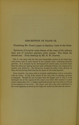
![SeprintMjrom ‘ Transactions of the Vatholoyical Society of London ’for 1882, *£> on. A*- fU( xxx/// J Ptyylka^cysts of the ovary. By Alban Doran. ' Jtoy // [With Plate IX.] The ovary is now known to be divided into two parts histologi- cally distinct, and differing in the manner of their develop- ment. Each part is subject to a different form of proliferous cyst. In discussing the nature of these specimens I will, in order to avoid confusing synonyms, employ the anatomical terms used by Dr. Klein and Mr. Noble Smith in their ‘Atlas of Histology.’ They speak of the Graafian follicles as the parenchyma. These follicles lie in the free, prominent part of the ovary, surrounded by the “ stroma of the parenchyma.”1 That portion of the ovary next to its attachment to the broad ligament they term the “ tissue of the hilum.”2 From the stroma of the parenchyma arise the simple as well as the multiple but not proliferous ovarian cysts,3 distinctly consisting of dilated Graafian follicles, ova being found in their cavities. From the same part arises the common multilocular proliferous cyst. From the inner walls of the parent, and secondary cysts spring, in many cases exuberant succulent growths, generally con- sidered to be glandular.4 The fluid contents are always glairy, and in the larger and older cysts more or less discoloured. The origin of these cysts is still disputed, whether they arise from the follicles at any stage of development or degeneration, or whether they owe their existence to changes beginning in the stroma or its vessels, it is not for me to discuss at present. As the seat of origin of these cysts is in the free part of the ovary, they rapidly absorb the structures in that part, so that when the tumour is only a few inches in diameter, the normal outline of the ovary is lost, and its healthy substance cannot at first be detected. As the hilum remains long free from disease, such cysts have a very distinct pedicle. 1 Syn.—Cortical portion, zone of Pfliiger’s (so-called) tubes. 3 %».—Medullary portion, zone of Kolliker’s “ Markstriinge des Hilus (sic) Ovarii.” 3 “ Oligocystic Tumours.” 4 Systematic German writers give to a cyst of this kind the lengthy but ex- pressive title, “ Cystoma ovarii proprium multiloculare proliferum glandulare.”](https://iiif.wellcomecollection.org/image/b22456703_0003.jp2/full/800%2C/0/default.jpg)