Distribution of histamine and substance P in the wall of the dog's digestive tract / by W.W. Douglas [and others].
- Date:
- [1951?]
Licence: In copyright
Credit: Distribution of histamine and substance P in the wall of the dog's digestive tract / by W.W. Douglas [and others]. Source: Wellcome Collection.
1/16
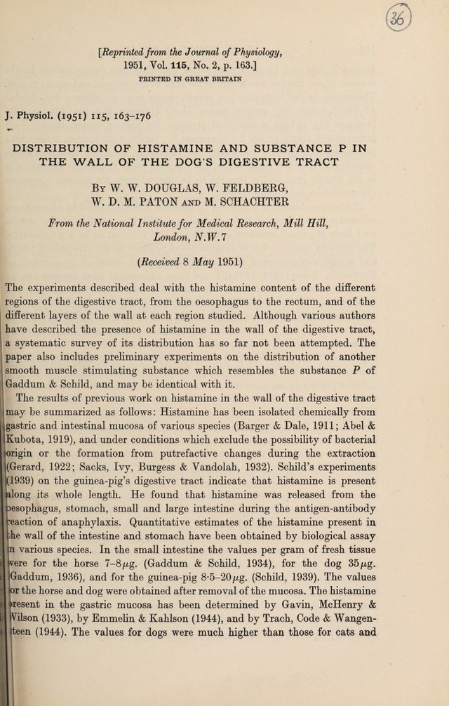
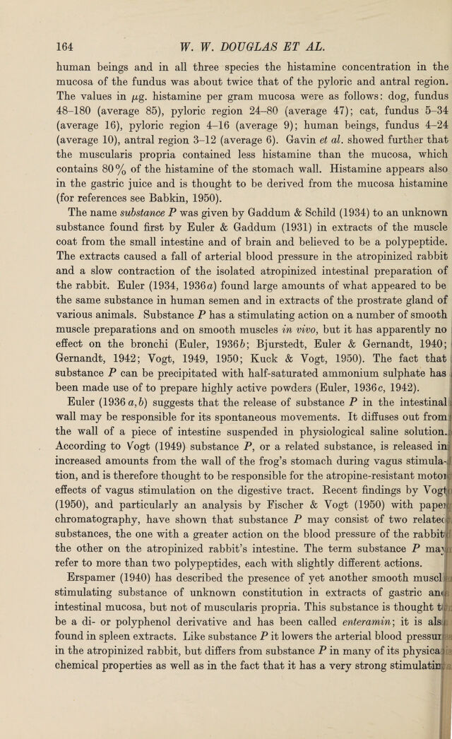
![[Reprinted from the Journal of Physiology, 1951, Yol. 115, No. 2, p. 163.] PRINTED IN GREAT BRITAIN J. Physiol. (1951) 115, 163-176 DISTRIBUTION OF HISTAMINE AND SUBSTANCE P IN THE WALL OF THE DOG’S DIGESTIVE TRACT By W. W. DOUGLAS, W. FELDBERG, W. D. M. PATON and M. SCHACHTER From the National Institute for Medical Research, Mill Hill, London, N.W.l [Received 8 May 1951) The experiments described deal with the histamine content of the different regions of the digestive tract, from the oesophagus to the rectum, and of the different layers of the wall at each region studied. Although various authors have described the presence of histamine in the wall of the digestive tract, a systematic survey of its distribution has so far not been attempted. The paper also includes preliminary experiments on the distribution of another smooth muscle stimulating substance which resembles the substance P of Gaddum & Schild, and may be identical with it. The results of previous work on histamine in the wall of the digestive tract may be summarized as follows: Histamine has been isolated chemically from jgastric and intestinal mucosa of various species (Barger & Dale, 1911; Abel & Kubota, 1919), and under conditions which exclude the possibility of bacterial origin or the formation from putrefactive changes during the extraction ‘(Gerard, 1922; Sacks, Ivy, Burgess & Yandolah, 1932). Schild’s experiments (1939) on the guinea-pig’s digestive tract indicate that histamine is present (along its whole length. He found that histamine was released from the oesophagus, stomach, small and large intestine during the antigen-antibody reaction of anaphylaxis. Quantitative estimates of the histamine present in j jphe wall of the intestine and stomach have been obtained by biological assay in various species. In the small intestine the values per gram of fresh tissue I (vere for the horse 7-8 pg. (Gaddum & Schild, 1934), for the dog 35 /xg. llGaddum, 1936), and for the guinea-pig 8*5-20jug. (Schild, 1939). The values or the horse and dog were obtained after removal of the mucosa. The histamine el present in the gastric mucosa has been determined by Gavin, McHenry & L iVilson (1933), by Emmelin & Kahlson (1944), and by Trach, Code & Wangen- >| feteen (1944). The values for dogs were much higher than those for cats and](https://iiif.wellcomecollection.org/image/b30633345_0001.jp2/full/800%2C/0/default.jpg)