Skip to main content
Wellcome Collection homepage
Visit us
What’s on
Stories
Collections
Get involved
About us
Sign in to your library account
Search for anything
Library account
Search for anything
Search
Home
|
Collections
Cranial Fossa, Posterior
Part of the cranial cavity, located between the foramen magnum and tentorium cerebelli
Wikidata
Source:
Wikidata
On this page
On this page
Images from the collections
Works from the collections
Related topics
Images from the collections
Images referencing Cranial Fossa, Posterior
3 images from works
Works from the collections
3 works
Digital Images
Online
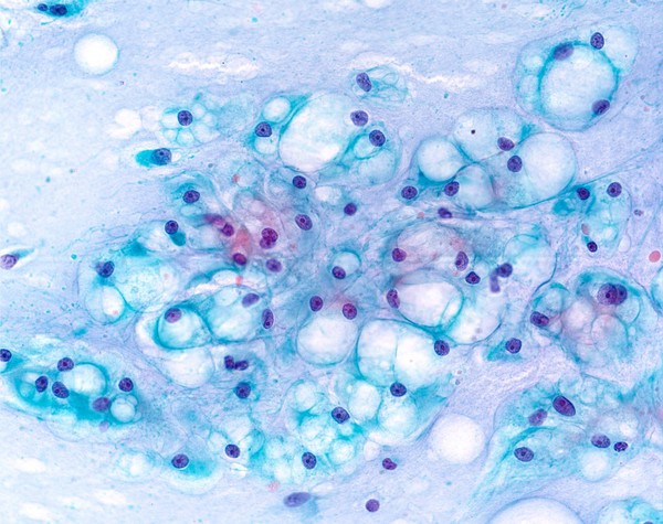
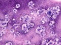
Papanicolaou stained smear of a clival chordoma, microscopy. Chordomas are cancers formed of cells which resemble those of the notochord (spine) of a developing foetus. Although they can present anywhere within the spine and skull, the majority grow in the sacral region of the spine, corresponding to the lower back. This image shows a Papanicolaou (Pap) stained smear obtained from a needle biopsy of a chordoma in the clivus, a part of the cranium at the base of the skull.
William R. Geddie
Digital Images
Online
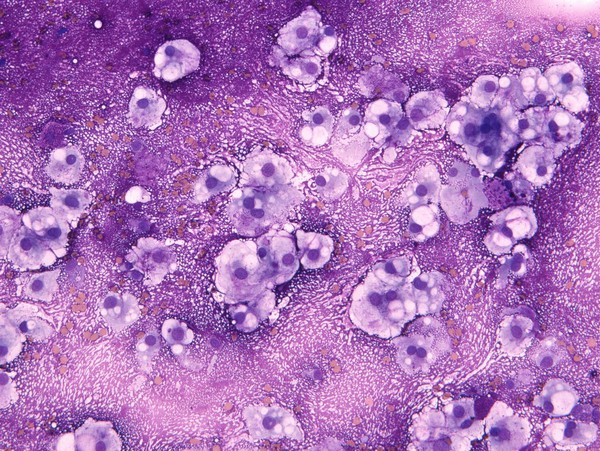
MGG stained smear of a C2 vertebral chordomal mass
William R. Geddie
Digital Images
Online
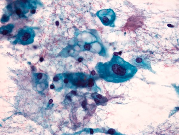
Papanicolaou stained smear of a C2 vertebral chordomal mass, microscopy. Chordomas are cancers formed of cells which resemble those of the notochord (spine) of a developing foetus. Although they can present anywhere within the spine and skull, the majority grow in the sacral region of the spine, corresponding to the lower back. This image shows a Papanicolaou (pap) stained smear obtained from a needle biopsy of a chordoma of the C2 vertebrae, located at the top of the neck just underneath the base of the skull.
William R. Geddie
Related topics
Purple
Oncology
Cancer
Skull
Skeleton
Bubbles
Smear
Mucous
Notocord
Cytology
Close modal window