Protection
Images from the collections
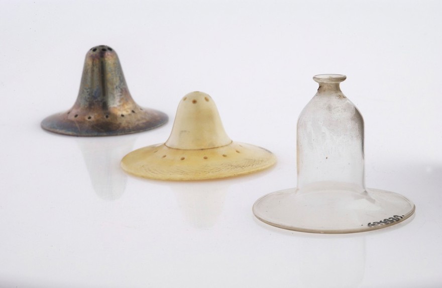

Works from the collections
46 works
- Digital Images
- Online
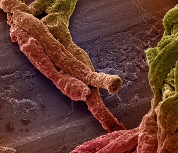
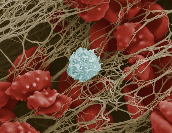
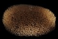
Cocoon from medicinal leech Hirudo verbena
Macroscopic Solutions
- Digital Images
- Online
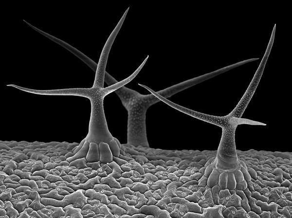
Stinging hairs on a nettle leaf
Liz Hirst, Medical Research Council
- Digital Images
- Online
Stinging nettle (Urtica dioica) stem, SEM
Kevin Mackenzie, University of Aberdeen
- Digital Images
- Online
Young girl with Down's syndrome
Fiona Yaron-Field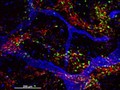
- Digital Images
- Online
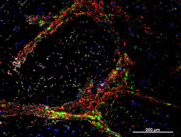
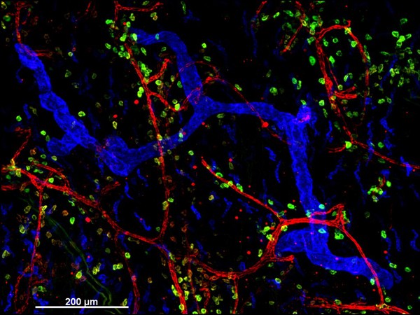
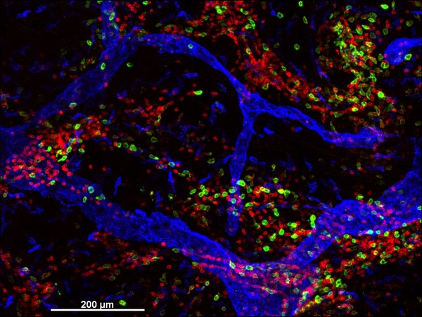
Cellular architecture of normal human skin imaged by whole mount tissue microscopy. Human skin has a rich network of white blood cells (specifically dendritic cells, T cells and macrophages) which form sheaths around blood vessels (string-like structures). A network of lymphatic vessels (ribbon-like structures) is also present. In this image, human skin lymphatic vessels (stained for LYVE-1; blue) and white blood cells comprised of dendritic cells (stained for CD11c; green) and T cells (stained for CD3; red) can be seen. Some macrophages also express the protein LYVE-1 similar to lymphatic vessel cells which can be appreciated as blue cells within and in between the sheaths of white blood cells. This normal cellular architecture is grossly disrupted in diseased skin (see related images). X10 magnification. Scale bar (white) represents 200 micrometres.
Dr. Xiao-nong Wang, Human Dendritic Cell Laboratory, Newcastle University