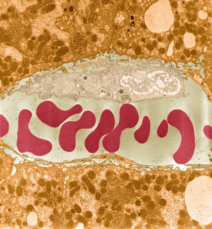
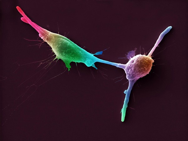
Leucocyte
Images from the collections
Works from the collections
30 works

- Digital Images
- Online
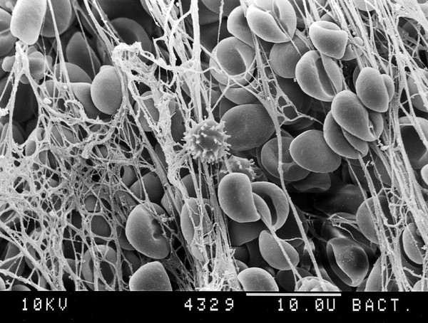
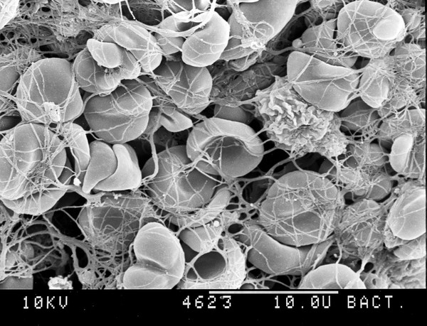
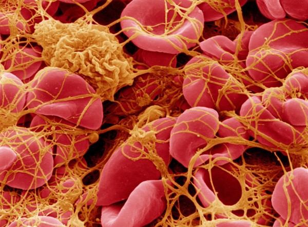
SEM of blood corpuscles in clot.
David Gregory & Debbie Marshall
- Digital Images
- Online
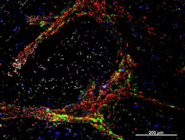
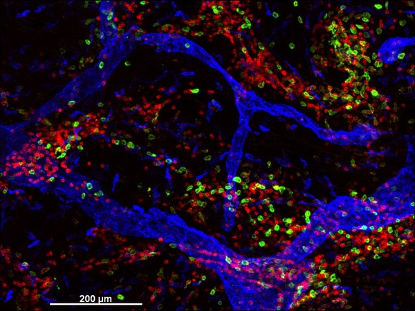
Castration resistant prostate cancer, human tissue
Mateus Crespo, The Institute of Cancer Research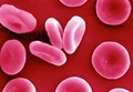
- Digital Images
- Online
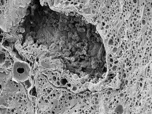
SEM of red blood corpuscles, close-up
David Gregory & Debbie Marshall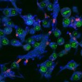
- Digital Images
- Online
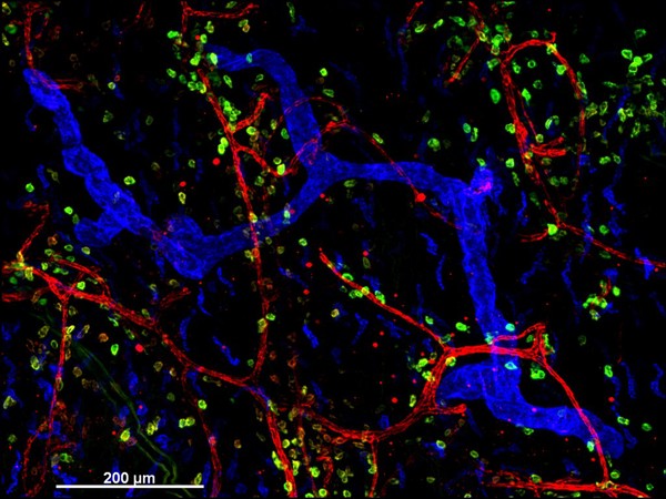
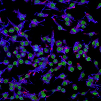
Macrophages infected with candida yeast, LM
Kevin Mackenzie, University of Aberdeen
- Digital Images
- Online
Macrophages infected with Candida yeast spores, TEM
Kevin Mackenzie, University of Aberdeen